Showing posts with label Lensless Camera. Show all posts

Saturday, June 04, 2016

Flat lens promises possible revolution in optics

http://www.bbc.com/news/science-environment-36438686

structure of the lens seen under microscopeImage copyrightFEDERICO CAPASSO
Image captionThis electron microscope image shows the structure of the lens (white line is 0.002mm long)
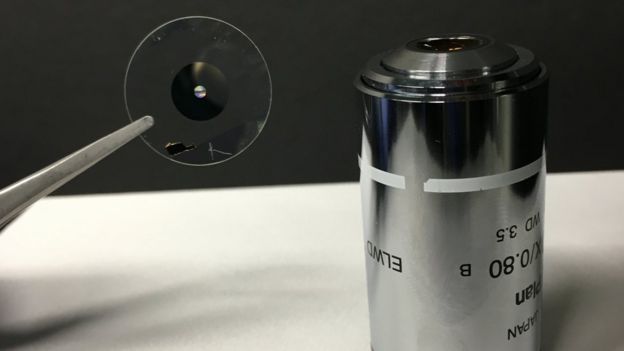
A flat lens made of paint whitener on a sliver of glass could revolutionise optics, according to its US inventors.
Just 2mm across and finer than a human hair, the tiny device can magnify nanoscale objects and gives a sharper focus than top-end microscope lenses.
It is the latest example of the power of metamaterials, whose novel properties emerge from their structure.
Shapes on the surface of this lens are smaller than the wavelength of light involved: a thousandth of a millimetre.
"In my opinion, this technology will be game-changing," said Federico Capasso of Harvard University, the senior author of a report on the new lens which appears in the journal Science.
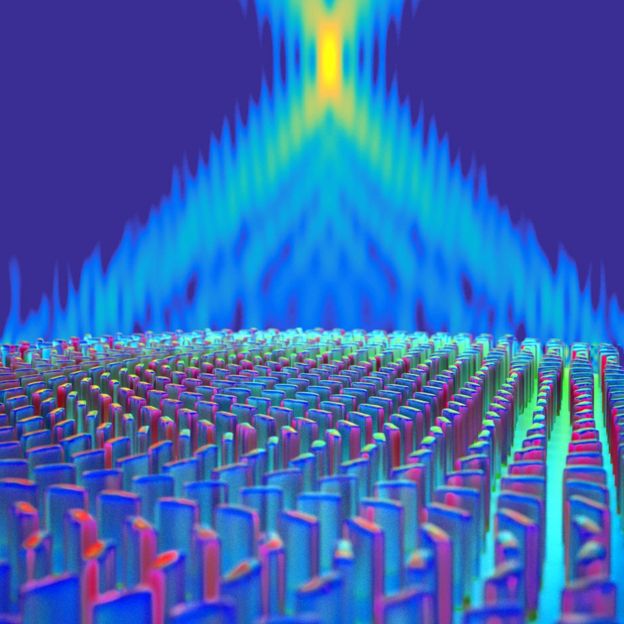
The lens is quite unlike the curved disks of glass familiar from cameras and binoculars. Instead, it is made of a thin layer of transparent quartz coated in millions of tiny pillars, each just tens of nanometres across and hundreds high.
Singly, each pillar interacts strongly with light. Their combined effect is to slice up a light beam and remould it as the rays pass through the array (see video below).
Media captionLight passing through the "metalens" is focussed by the array of nanostructures on its surface (video: Capasso Lab/Harvard)
Computer calculations are needed to find the exact pattern which will replicate the focussing effect of a conventional lens.
The advantage, Prof Capasso said, is that these "metalenses" avoid shortfalls - called aberrations - that are inherent in traditional glass optics.
"The quality of our images is actually better than with a state-of-the-art objective lens. I think it is no exaggeration to say that this is potentially revolutionary."
Those comparisons were made against top-end lenses used in research microscopes, designed to achieve absolute maximum magnification. The focal spot of the flat lens was typically 30% sharper than its competition, meaning that in a lab setting, finer details can be revealed.
But the technology could be revolutionary for another reason, Prof Capasso maintains.
"The conventional fabrication of shaped lenses depends on moulding and essentially goes back to 19th Century technology.
"But our lenses, being planar, can be fabricated in the same foundries that make computer chips. So all of a sudden the factories that make integrated circuits can make our lenses."
And with ease. Electronics manufacturers making microprocessors and memory chips routinely craft components far smaller than the pillars in the flat lenses. Yet a memory chip containing billions of components may cost just a few pounds.
two lenses side by sideImage copyrightFEDERICO CAPASSO
Image captionThe lens is much more compact than a traditional microscope objective
Mass production is the key to managing costs, which is why Prof Capasso sees cell-phone cameras as an obvious target. Most of their other components, including the camera's detector, are already made with chip technology. Extending that to include the lens would be natural, he argues.
There are many other potential uses: mass-produced cameras for quality control in factories, light-weight optics for virtual-reality headsets, even contact lenses. "We can make these on soft materials," Prof Capasso assured the BBC.
The prototypes lenses are 2mm across, but only because of the limitations of the Harvard manufacturing equipment. In principle, the method could scale to any size, Prof Capasso said.
"Once you have the foundry - you want a 12-inch lens? Feel free, you can make a 12-inch lens. There's no limit."
The precise character of the lens depends on the layout and composition of the pillars. Paint-whitener - titanium dioxide - is used to make the pillars, because it is transparent and interacts strongly with visible light. It is also cheap.
illustration of light hitting the lensImage copyrightPETER ALLEN/HARVARD
Image captionThe minuscule pillars have a powerful effect on light passing through
The team has previously worked with silicon, which functions well in the infrared. Other materials could be used to make ultraviolet lenses.
Or to get a different focus, engineers could change the size, spacing and orientation of the pillars. It simply means doing the computer calculations and dialling the results into the new design.
The team is already working on beating the performance of its first prototypes. Watch this space, they say - if possible, with a pair of metalenses.

Tuesday, February 10, 2015

Friday, December 26, 2014

lensfree holographic on-chip microscopy


Actually this shouldn't be that hard to do.  It is computational photography at it's finest.

It should be able to completely put to shame normal optical microscopes.
It is volumetric and 3D viewable and could even go multi-spectral. 

There's no information on the specifics of the optics, but the sample must go directly on the imaging chip or very close to it.

So cleaning and reuse are my only questions.

Lens-free microscope can detect cancer at the cellular level

UCLA researchers develop device that can do the work of pathology lab microscopes

http://newsroom.ucla.edu/releases/lens-free-microscope-can-detect-cancer-at-the-cellular-level


 The latest invention is the first lens-free microscope that can be used for high-throughput 3-D tissue imaging — an important need in the study of disease.

“This is a milestone in the work we’ve been doing,” said Ozcan, who also is the associate director of UCLA’s California NanoSystems Institute. “This is the first time tissue samples have been imaged in 3D using a lens-free on-chip microscope.”

The device works by using a laser or light-emitting-diode to illuminate a tissue or blood sample that has been placed on a slide and inserted into the device. A sensor array on a microchip — the same type of chip that is used in digital cameras, including cellphone cameras — captures and records the pattern of shadows created by the sample.

The device processes these patterns as a series of holograms, forming 3-D images of the specimen and giving medical personnel a virtual depth-of-field view. An algorithm color codes the reconstructed images, making the contrasts in the samples more apparent than they would be in the holograms and making any abnormalities easier to detect.


Wide-field computational imaging of pathology slides using lens-free on-chip microscopy


Alon Greenbaum, Yibo Zhang,  Alborz Feizi, Ping-Luen Chung, Wei Luo, Shivani R. Kandukuri and Aydogan Ozcan

http://stm.sciencemag.org/content/6/267/267ra175

Optical examination of microscale features in pathology slides is one of the gold standards to diagnose disease. However, the use of conventional light microscopes is partially limited owing to their relatively high cost, bulkiness of lens-based optics, small field of view (FOV), and requirements for lateral scanning and three-dimensional (3D) focus adjustment. We illustrate the performance of a computational lens-free, holographic on-chip microscope that uses the transport-of-intensity equation, multi-height iterative phase retrieval, and rotational field transformations to perform wide-FOV imaging of pathology samples with comparable image quality to a traditional transmission lens-based microscope. The holographically reconstructed image can be digitally focused at any depth within the object FOV (after image capture) without the need for mechanical focus adjustment and is also digitally corrected for artifacts arising from uncontrolled tilting and height variations between the sample and sensor planes. Using this lens-free on-chip microscope, we successfully imaged invasive carcinoma cells within human breast sections, Papanicolaou smears revealing a high-grade squamous intraepithelial lesion, and sickle cell anemia blood smears over a FOV of 20.5 mm2. The resulting wide-field lens-free images had sufficient image resolution and contrast for clinical evaluation, as demonstrated by a pathologist’s blinded diagnosis of breast cancer tissue samples, achieving an overall accuracy of ~99%. By providing high-resolution images of large-area pathology samples with 3D digital focus adjustment, lens-free on-chip microscopy can be useful in resource-limited and point-of-care settings.

Toward giga-pixel nanoscopy on a chip: a computational wide-field look at the nano-scale without the use of lenses


http://pubs.rsc.org/en/content/articlelanding/2013/lc/c3lc50222h#!divAbstract


The development of lensfree on-chip microscopy in the past decade has opened up various new possibilities for biomedical imaging across ultra-large fields of view using compact, portable, and cost-effective devices. However, until recently, its ability to resolve fine features and detect ultra-small particles has not rivalled the capabilities of the more expensive and bulky laboratory-grade optical microscopes. In this Frontier Review, we highlight the developments over the last two years that have enabled computational lensfree holographic on-chip microscopy to compete with and, in some cases, surpass conventional bright-field microscopy in its ability to image nano-scale objects across large fields of view, yielding giga-pixel phase and amplitude images. Lensfree microscopy has now achieved a numerical aperture as high as 0.92, with a spatial resolution as small as 225 nm across a large field of view e.g., >20 mm2. Furthermore, the combination of lensfree microscopy with self-assembled nanolenses, forming nano-catenoid minimal surfaces around individual nanoparticles has boosted the image contrast to levels high enough to permit bright-field imaging of individual particles smaller than 100 nm. These capabilities support a number of new applications, including, for example, the detection and sizing of individual virus particles using field-portable computational on-chip microscopes.

Thursday, December 04, 2014

Rambus Lensless Camera Demo

From: http://image-sensors-world.blogspot.com/2014/12/rambus-lensless-camera-demo.html

Rambus publishes a Youtube video with Patrick Gill showing the company's lensless camera operation:

Dr. Patrick Gill demonstrates a diffraction-based lensless imaging system

 Meanwhile, it appears that Rambus somewhat downplays its image sensor activities in its recent investor presentations. For example, in the Nov. 2014 presentation, imaging appears in only one slide #29:

Tuesday, June 04, 2013

Bell Labs creates a lensless camera that's always in focus


Bell Labs creates a lensless camera that's always in focus


One day, cameras may be able to capture less data but produce images that look just as good as a traditional photo. Bell Labs is the latest to attempt such a feat, and it's doing so while eschewing another major camera standby, the lens. The laboratory has developed a single-pixel camera that only uses a series of transparent openings to capture its image, without any glass to direct the light. The system uses a technology called "compressive sensing," which is still in its early stages of study. The idea is that instead of a camera capturing a full image and then whittling the data down into a small, compressed file like a JPG, a camera could instead capture almost exactly what it needs, making capture times much quicker.
NO LENS, ALWAYS IN FOCUS
This isn't a reality just yet, however. Compressive sensing cameras build their final image by comparing the differences that come in through each aperture, and right now that takes too much time for them to shoot anything other than a still life. But by removing the lens, Bell Labs adds another impressive feature to its camera: its shots are always in focus. One always-in-focus camera, the Lytro, is already on the market, but Bell Labs sees its new tech as a practical way to shrink the size and cost of future cameras.

Bell Labs' device is built with "low cost, commercially available components," which primarily amount to a semi-transparent LCD panel, a one-megapixel imaging sensor, and a computer to connect it all to. The LCD panel was placed in front of the sensor, and light came in through white "openings" in the panel. The camera measured the data separately for red, green, and blue light, and used a computer to stitch together the final image. While the images don't demonstrate the finest image quality, they emphasize what compressive sensing is capable of. The books were captured using only a quarter of the camera's total imaging capabilities, and the soccer ball was captured using even less, just one-eighth.